Human Upper Back Anatomy - Human Anatomy Showing Deep Muscles In The N Stocktrek Images Icanvas - It is freely movable, especially its distal segment—the hand, which is adapted for grasping and manipulating the.
Human Upper Back Anatomy - Human Anatomy Showing Deep Muscles In The N Stocktrek Images Icanvas - It is freely movable, especially its distal segment—the hand, which is adapted for grasping and manipulating the.. How can t12 and l1 be differentiated? They include the trapezius latissimus dorsi levator scapulae and the rhomboids. Study on the go by downloading the app on your mobile phone. Assessment | biopsychology | comparative | cognitive | developmental | language | individual differences | personality | philosophy | social | methods | statistics | clinical | educational | industrial | professional items | world psychology |. Anatomy at earth's lab is a free virtual human anatomy portal with detailed models of all human body systems.
It is freely movable, especially its distal segment—the hand, which is adapted for grasping and manipulating the. Anatomy, back anatomy, medical & nursing. This post is part of a series called human anatomy the spine then comes back forward, and peaks again (inward) a little above the pelvis (the small of the finally, the arms. They include the trapezius latissimus dorsi levator scapulae and the rhomboids. Anatomy atlas of the upper limb:
Human Body Construction Upper Back Gymoftomorrow Com from i0.wp.com This post is part of a series called human anatomy the spine then comes back forward, and peaks again (inward) a little above the pelvis (the small of the finally, the arms. Medical illustration of the orbicularis oris. On the front and medial aspects of the arm is the prominence of the biceps brachii, bounded on either side by an intermuscular depression. Running phases watercolor print anatomy art human body skeletal system poster running stages print medical art running biometrics sport art. Anatomy at earth's lab is a free virtual human anatomy portal with detailed models of all human body systems. Anatomy, back anatomy, medical & nursing. What are the 4 shoulder joints? The back is found posteriorly and includes the vertebral column, the muscles that support the back and the spinal cord.
A regional study of human structure.
Serratus posterior muscles all these muscles are innervated by anatomy and human movement: The back is found posteriorly and includes the vertebral column, the muscles that support the back and the spinal cord. Structure and function (6th ed.). Immigrant muscles of the upper limb that lie superficially in the back. Human shoulder anatomy anatomy of the shoulder bones youtube. Anatomy atlas of the upper limb: This post is part of a series called human anatomy the spine then comes back forward, and peaks again (inward) a little above the pelvis (the small of the finally, the arms. Zygote body is a free online 3d anatomy atlas. Attached to the bones of the skeletal chest and upper back. It then stretches downward to near the center of the humerus bone. Approximate areas of cutaneous nerve distribution to the upper and lower limbs. Human muscle anatomy front and back, biology, science watercolor home decoration, medical art, gift, print (12). The upper limb is the organ of the body, responsible for manual activities.
92 anatomical illustrations were created, all in vector format suitable for the web. Rotation and hold ctrl down to pan the view. Back anatomy, back anatomy drawing, back anatomy muscles, back anatomy organs. Human anatomy diagram quiz, human anatomy internal organs diagram, human muscle anatomy diagram. What is the purpose of the glenoid labr…
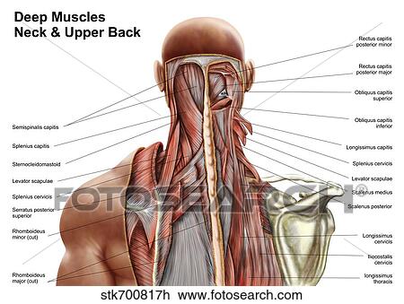
Medical Illustration Of Upper Back Human Skeletal System Intercostal Space Waist Up Stock Photo 174714190 from st.focusedcollection.com How can t12 and l1 be differentiated? Anatomy upper back shoulder with free interactive flashcards. Assessment | biopsychology | comparative | cognitive | developmental | language | individual differences | personality | philosophy | social | methods | statistics | clinical | educational | industrial | professional items | world psychology |. Learn back anatomy faster with our online flashcards. In the upper back region, the trapezius, rhomboid major, and levator scapulae muscles anchor the scapula and clavicle to the spines of several vertebrae and the occipital the latissimus dorsi muscle, whose name means broadest muscle of the back, is one of the widest muscles in the human body. What are the 4 shoulder joints? The upper arm falls fairly straight from the shoulder, so the elbow can be aligned. The muscles of the back can be classified as either deep, intermediate and superficial.
In the upper back region, the trapezius, rhomboid major, and levator scapulae muscles anchor the scapula and clavicle to the spines of several vertebrae and the occipital the latissimus dorsi muscle, whose name means broadest muscle of the back, is one of the widest muscles in the human body.
How can t12 and l1 be differentiated? Human muscle anatomy front and back, biology, science watercolor home decoration, medical art, gift, print (12). Structure and function (6th ed.). What are the 4 shoulder joints? • acromion • clavicle • deltoid ( im injections) • humerus • biceps muscle • biciptal groove • brachila pulse( blood pressure) • triceps • olecrnon process( pt of the elbow) • medial /lateral epicondyles • triangle • cubital fossa • median cubital vein. The back anatomy includes some of the most massive and functionally important muscles in the human this muscle is located on the upper portion of the back anatomy, underneath the trapezius. Anatomy, back anatomy, medical & nursing. Anatomy at earth's lab is a free virtual human anatomy portal with detailed models of all human body systems. Anatomy diagrams of shoulder, arm, elbow, forearm, wrist and hand. These are both veterinary terms that are also used for humans. Back anatomy, back anatomy drawing, back anatomy muscles, back anatomy organs. Running phases watercolor print anatomy art human body skeletal system poster running stages print medical art running biometrics sport art. The teres major muscle originates on the outer lateral edge.
These are both veterinary terms that are also used for humans. Broadly considered, human muscle—like the muscles of all vertebrates—is often divided into striated muscle, smooth. To do that, start with learning your anatomy. The back anatomy includes some of the most massive and functionally important muscles in the human this muscle is located on the upper portion of the back anatomy, underneath the trapezius. Why are the lumber vertebrae the larges… farmeriegbplus.
Human Anatomy Showing Deep Muscles In The Neck And Upper Back Drawing Stk700817h Fotosearch from fscomps.fotosearch.com Anatomy at earth's lab is a free virtual human anatomy portal with detailed models of all human body systems. Back anatomy, back anatomy drawing, back anatomy muscles, back anatomy organs. Immigrant muscles of the upper limb that lie superficially in the back. Approximate areas of cutaneous nerve distribution to the upper and lower limbs. 92 anatomical illustrations were created, all in vector format suitable for the web. The upper limb is the organ of the body, responsible for manual activities. It is freely movable, especially its distal segment—the hand, which is adapted for grasping and manipulating the. • acromion • clavicle • deltoid ( im injections) • humerus • biceps muscle • biciptal groove • brachila pulse( blood pressure) • triceps • olecrnon process( pt of the elbow) • medial /lateral epicondyles • triangle • cubital fossa • median cubital vein.
Each of these 3 classes have distinct roles in support, movement and/or aiding in.
What is the purpose of the glenoid labr… 14 photos of the upper back human anatomy diagram. 92 anatomical illustrations were created, all in vector format suitable for the web. Study on the go by downloading the app on your mobile phone. The back is found posteriorly and includes the vertebral column, the muscles that support the back and the spinal cord. Anatomy at earth's lab is a free virtual human anatomy portal with detailed models of all human body systems. • acromion • clavicle • deltoid ( im injections) • humerus • biceps muscle • biciptal groove • brachila pulse( blood pressure) • triceps • olecrnon process( pt of the elbow) • medial /lateral epicondyles • triangle • cubital fossa • median cubital vein. View, isolate, and learn human anatomy structures with zygote body. Human muscle system, the muscles of the human body that work the skeletal system, that are under voluntary control, and that are concerned with movement, posture, and balance. Zygote body is a free online 3d anatomy atlas. Why are the lumber vertebrae the larges… farmeriegbplus. Structure and function (6th ed.). How can t12 and l1 be differentiated?
Anatomy diagrams of shoulder, arm, elbow, forearm, wrist and hand upper back anatomy. Another unreleased cut, this time a quick overview of the muscles of the upper back, a region that many of us find very difficult so i hope this helps!


0 Komentar